
LESSON 3 – ECG Waveforms
- If the amplitude exceeds 5 mm, we use uppercase letters (Q, R, or S).
- If the amplitude is 5 mm or less, we use lowercase letters (q, r, or s).
- Note: This rule does not apply to P or T waves.
- Q and S waves are always negative (below the isoelectric line).
- R waves are always positive (above the isoelectric line).
- P and T waves can be either positive or negative depending on the lead and clinical context.
Let’s look at an example for clarity.
We will take a sample complex and define the isoelectric line—the segment of the tracing immediately following the T wave where no electrical activity is recorded.”
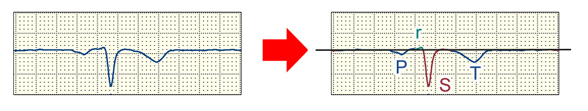
Now, simply put: everything above the isoelectric line is a positive deflection, and everything below it is a negative deflection.
Looking at our first example: the first deflection after the baseline is a P wave, and in this specific case, it is negative. Next, we see a positive deflection. Since this is the first upward movement, it must be an R wave. However, its amplitude is only 1 mm (less than 5 mm), so we designate it with a lowercase ‘r’. Following the ‘r’ wave, a negative deflection appears—this is the S wave (recall that any negative wave following an R is an S). Its amplitude exceeds 5 mm, so it earns an uppercase ‘S’. Finally, we have the T wave, which is also negative here.
Is it all clear? Well, perhaps not entirely! You might ask: ‘Where is the Q wave?’ In this instance, it’s absent—which is a common finding. By definition, a Q wave is the first negative deflection following the P wave but preceding the R wave. Since our complex began with an ‘r’ wave, there is no Q. Now, the picture should be getting clearer: if someone asks about the morphology of this ventricular complex, you can confidently call it an ‘rS complex.’
Let’s examine another complex! Again, we draw our isoelectric line and observe the deflections. In this case, the P wave is absent. While this is an abnormality (not a normal sinus rhythm), let’s set that aside for now to focus purely on the nomenclature. This tracing begins directly with the ventricular complex…”
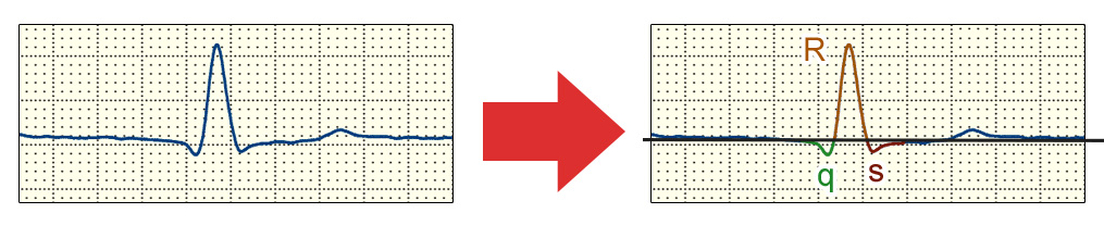
Following the isoelectric line, a negative deflection appears—this is a q wave (lowercase ‘q’ because its amplitude is less than 5 mm). Next, a tall R wave emerges (uppercase ‘R’ because it exceeds 5 mm), followed by another negative deflection—an s wave (lowercase ‘s’ because its amplitude is less than 5 mm). Thus, the morphology of this ventricular complex is defined as ‘qRs’. Not too difficult, right? However… what if we encounter something like this…”
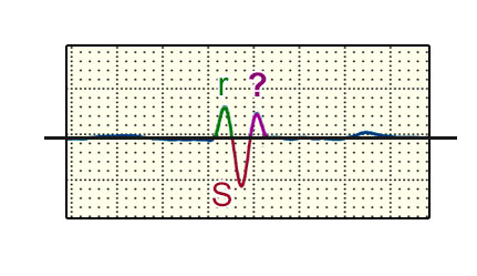
At first glance, it starts predictably: an rS… but what follows? It’s actually quite simple — if a second ‘r’ wave appears, we simply denote it with a ‘prime’ symbol: r’. Therefore, the morphology of this specific ventricular complex is rSr’.
From now on, whenever you encounter these notations in medical literature, you will know exactly what they describe.
To solidify this knowledge, let’s move on to a very simple exercise…