
LESSON 6 – How to Assess Proper ECG Recording
We all make mistakes from time to time. During ECG recording, electrode misplacement is not uncommon. If one or two precordial electrodes are misplaced, it is usually not a major problem, as the ECG typically does not change significantly. However, if the right and left arm electrodes or leg electrodes are interchanged—which happens much more often—it can create significant diagnostic confusion and may even mimic myocardial infarction.
So how can you avoid this pitfall?
The answer lies in the aVR lead. If in lead aVR the P wave and T wave are positive, and there is also a tall positive R wave, then with a probability of 99.9% the limb electrodes have been misplaced.
Such an ECG must not be interpreted. The recording should be repeated with correct electrode placement, and only then should the ECG be analyzed.
Example of an ECG with misplaced electrodes
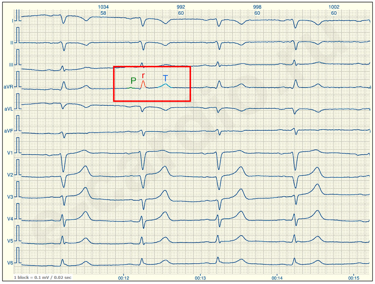
Explanation:
In lead aVR, the P wave and T wave are positive, and a tall R wave is present.
ECG of the same patient with correct electrode placement
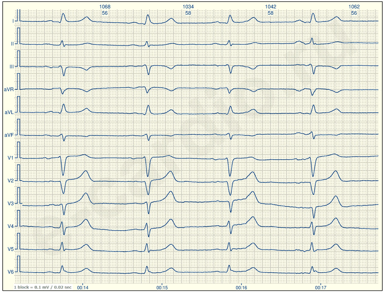
Explanation:
In lead aVR, as normally expected (check your reference sheet), the P wave and T wave are negative. The R wave in lead aVR is almost completely absent.
Not difficult, right?
But to check how well you understood this concept, I have prepared a small but tricky task for you 🙂.