
LESSON 11 – Wandering atrial pacemaker and Sinus arrhythmia
This rhythm represents a combination of sinus and supraventricular rhythms alternating from beat to beat. The P waves continuously change their morphology, polarity, and their relationship to the QRS complex. However, each P wave is followed by a QRS complex.
Let’s go straight to an example. I deliberately chose an ECG without a pronounced wandering pattern (not the classic textbook appearance). Therefore, I would like to immediately draw your attention to leads aVF and II — these are the primary leads where atrial activity should always be assessed first.
Example of an ECG with wandering atrial pacemaker
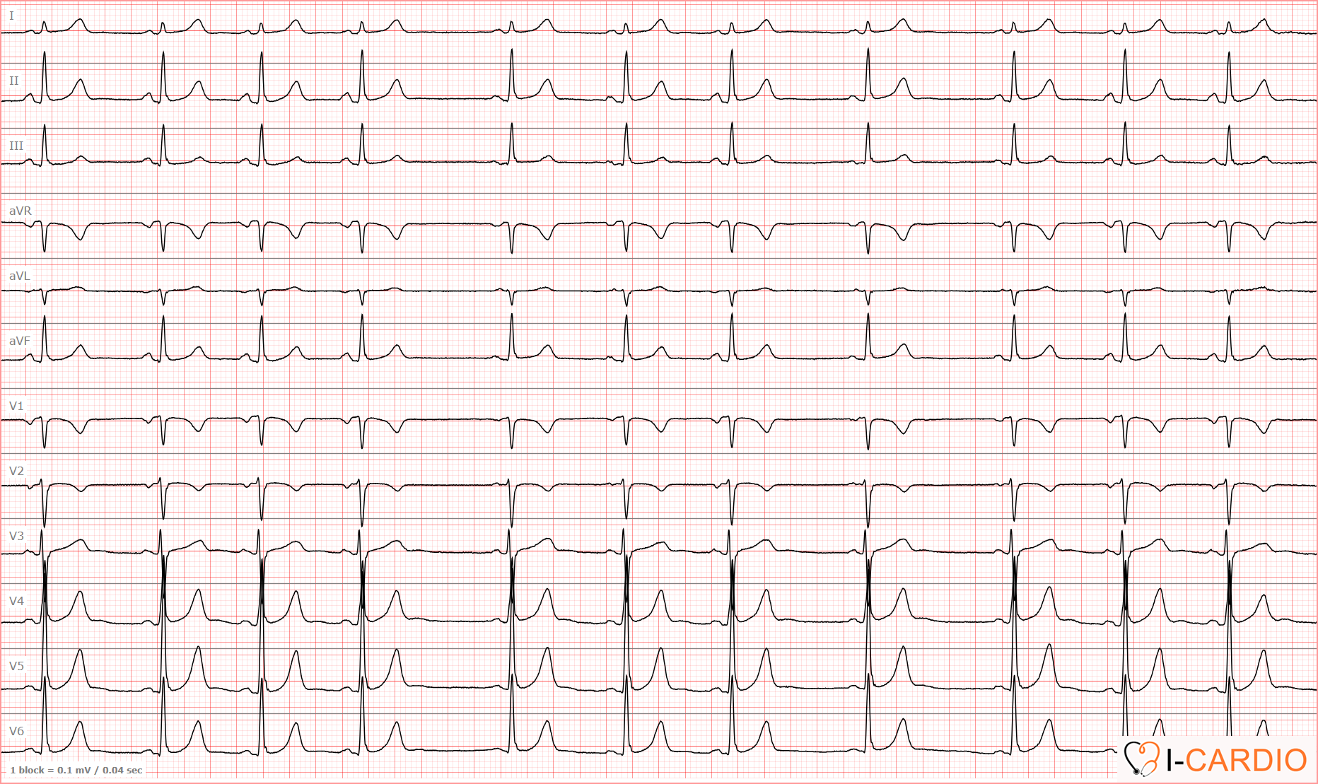
This ECG shows a mildly pronounced wandering atrial pacemaker. The first four P waves are likely sinus in origin. However, P waves “5” and “6” differ not only in morphology but also in their PR intervals. P wave “7” resembles the initial complexes, whereas “8” again differs. P wave “9” appears intermediate, while “10” and “11” are identical to the first four.

In addition to P-wave morphology, the duration of the PQ (PR) interval is critically important for diagnosis! If the PQ (PR) intervals remain unchanged while the P-wave morphology varies only slightly, this is usually not due to pacemaker migration, but rather to technical factors or transient changes in intra-atrial conduction.

A wandering atrial pacemaker generally has little clinical significance and is more commonly observed in young, healthy individuals.
However, it is important to be familiar with this type of rhythm disturbance and be able to recognize it in order not to confuse it with more serious arrhythmias. In addition, ECG recordings with multiple artifacts (baseline wander, electrical interference, etc.) may mimic this phenomenon.
Sinus arrhythmia
When discussing a wandering atrial pacemaker, it is also essential to address sinus arrhythmia.
Sinus arrhythmia on ECG is an irregular heart rhythm in which the normal impulse origin (the sinus node) is preserved, but the intervals between beats (RR intervals) vary by more than 0.12 seconds and/or more than 10%.
It is commonly observed in children, adolescents, and athletes, is often related to respiration (heart rate increases during inspiration), and is generally considered a physiological variant.
In other words, the criteria for this rhythm are identical to those of sinus rhythm, except for the final parameter — heart rate variability.
Criteria for Sinus Arrhyrmia
- In lead II, and usually also in leads I and aVF, there are positive P waves of identical morphology, located at a constant distance from the QRS complex in all consecutive cardiac cycles.
- The number of P waves equals the number of QRS complexes.
- Heart rate is between 60 and 100 beats per minute. Values below this range indicate sinus bradycardia, and values above it indicate sinus tachycardia.
- RR intervals are regular: variation between consecutive RR intervals > 10% of the mean RR interval duration, or > 120 ms.
Example of an ECG with sinus arrhythmia
Let us analyze lead II.
All criteria of normal sinus rhythm are present, except for variability.
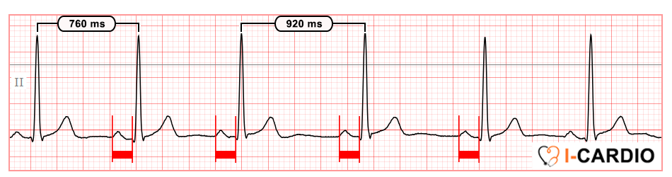
Difference between RR intervals: 920 ms − 760 ms = 160 ms (normal value < 120 ms).
Mean RR interval: (920 ms + 760 ms) / 2 = 840 ms. RR variability: (160 / 840) × 100 ≈ 19% (normal value ≤ 10%).
In contrast to a wandering pacemaker, in sinus arrhythmia the P-wave morphology and the PR interval remain constant, this is a key distinguishing feature. Although both conditions are characterized by significant heart rate variability.
Now let’s reinforce what we’ve learned with a practical task!